The field of medical imaging has undergone remarkable advancements in recent years, offering clinicians an invaluable tool for understanding and diagnosing various conditions. Among the most commonly used imaging techniques, computed tomography (CT) has become a cornerstone in clinical practice, especially in the evaluation of head and neck anatomy. With its ability to provide detailed cross-sectional images, CT scanning allows for a precise evaluation of the structures within these regions. One of the most effective ways of enhancing the utility of CT imaging in this context is through sagittal labeling, which offers specific insights into the anatomy of the head and neck. This article explores the intersection of anatomy and imaging with a focus on CT head and neck sagittal labeling, highlighting its importance and the insights it provides in both clinical and educational settings.
The Role of CT Imaging in Head and Neck Anatomy
CT imaging has revolutionized the way clinicians view the complex anatomy of the head and neck. The head and neck region consists of a wide variety of structures, including the brain, spinal cord, blood vessels, nerves, muscles, and soft tissues, which are often challenging to visualize using conventional techniques like X-ray. CT scans provide detailed, high-resolution images that allow for the visualization of both bone and soft tissue, making them an essential tool for diagnosis and treatment planning in this area.
CT scans use a series of X-ray images taken from different angles, which are then processed to create cross-sectional images or “slices” of the body. These slices provide clinicians with a clear view of internal structures, aiding in the identification of abnormalities such as tumors, fractures, infections, and other pathologies. In the head and neck region, CT imaging is particularly useful in assessing conditions such as cancer, trauma, vascular abnormalities, and neurological disorders.
Sagittal Plane: An Overview
To fully appreciate the value of sagittal labeling in CT imaging, it is essential to understand the significance of the sagittal plane in anatomical imaging. The body can be divided into three main planes: the sagittal, coronal, and transverse planes.
-
Sagittal Plane: The sagittal plane divides the body into left and right halves. It is a vertical plane that runs parallel to the body’s midline, offering a side view of the body and its structures.
-
Coronal Plane: The coronal plane divides the body into front and back halves, providing a view of the body from the front or posterior.
-
Transverse Plane: The transverse plane divides the body into top and bottom halves, offering a cross-sectional view from above or below.
The sagittal plane is particularly useful in imaging the head and neck because it offers a lateral view of these regions. This plane is crucial for evaluating structures such as the brain, spine, nasal cavity, pharynx, larynx, and major blood vessels. CT sagittal imaging provides a unique perspective that aids in understanding the spatial relationships between these complex structures.
Importance of Sagittal Labeling in CT Imaging
Sagittal labeling involves marking the key anatomical structures on CT images, allowing for easier identification and interpretation. In the context of head and neck imaging, this process is crucial for several reasons:
-
Improved Localization: Labeling allows clinicians to precisely locate anatomical structures within the sagittal plane. This is especially important for evaluating complex structures like the cervical spine, brainstem, and vascular systems. For example, labeling can clearly delineate the position of the carotid artery relative to the vertebral column, which is vital for surgical planning or understanding the spread of pathology.
-
Enhanced Diagnosis and Treatment Planning: When tumors, fractures, or other pathological conditions are identified, sagittal labeling allows clinicians to assess the extent and location of the issue with greater accuracy. For example, in cases of head and neck cancer, knowing the precise relationship between a tumor and surrounding structures, such as the lymph nodes, is critical for planning surgery or radiation therapy.
-
Educational Value: Sagittal labeling is also invaluable in medical education. It allows students and healthcare professionals to better understand the anatomical relationships and variations within the head and neck region. By labeling structures such as the pharyngeal muscles, cranial nerves, and sinuses, educators can enhance their students’ ability to interpret CT images and improve their anatomical knowledge.
-
Facilitating Surgical Procedures: In surgery, particularly in complex head and neck surgeries, having a clear understanding of anatomical relationships in the sagittal plane can reduce the risk of complications. For example, surgeons performing a cervical spine procedure can use sagittal images to navigate the delicate structures of the spinal cord and surrounding tissues. Similarly, otolaryngologists can use sagittal views to plan approaches to the sinuses or the larynx.
Insights from CT Head and Neck Sagittal Labeling
CT imaging with sagittal labeling provides multiple valuable insights into the anatomy of the head and neck region. Below are some key insights obtained from this form of imaging:
-
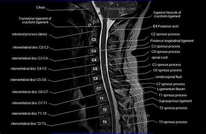
Cervical Spine and Spinal Cord: The sagittal view of the cervical spine allows clinicians to assess alignment, curvature, and any abnormalities such as disc herniations or fractures. In trauma cases, sagittal labeling can help identify vertebral fractures and the impact of trauma on the spinal cord, potentially guiding interventions such as surgery or immobilization.
-
Brain and Brainstem: The sagittal plane is essential for understanding the anatomy of the brain and brainstem. CT images can highlight various parts of the brain, such as the cerebellum, pons, and medulla. This is particularly important for detecting conditions like brain tumors, hemorrhages, and congenital anomalies.
-
Pharyngeal and Laryngeal Structures: The sagittal view also provides valuable information about the pharynx and larynx, essential for diagnosing conditions like obstructions, tumors, or swallowing disorders. These insights are particularly beneficial for otolaryngologists and speech therapists.
-
Sinuses and Nasal Cavity: Detailed labeling of the nasal passages and sinuses in the sagittal plane helps identify sinusitis, polyps, or other obstructive conditions. This is critical in planning surgical interventions such as sinus surgeries or septoplasty.
-
Vascular Structures: The major blood vessels of the head and neck, such as the carotid arteries and jugular veins, are easily identifiable in sagittal CT images. Sagittal labeling helps to evaluate the course of these vessels, identify stenosis or aneurysms, and plan surgeries or interventions like stent placement.
Conclusion
The intersection of anatomy and imaging is a critical area of study in modern medicine. CT head and neck sagittal labeling plays an essential role in this intersection by providing detailed and accurate insights into the complex structures of the head and neck. From diagnostic purposes to surgical planning, this imaging technique enhances the clinician’s ability to make informed decisions, ensuring better patient outcomes. Additionally, the educational value of sagittal labeling cannot be overstated, as it helps train medical professionals to interpret complex anatomical relationships. As medical imaging technology continues to evolve, the role of CT scans in providing deeper insights into human anatomy will only become more significant, offering new opportunities for diagnosis, treatment, and patient care. Visit Health Dady to get more information.